Copyright
©2013 Baishideng Publishing Group Co.
World J Gastroenterol. Nov 14, 2013; 19(42): 7241-7246
Published online Nov 14, 2013. doi: 10.3748/wjg.v19.i42.7241
Published online Nov 14, 2013. doi: 10.3748/wjg.v19.i42.7241
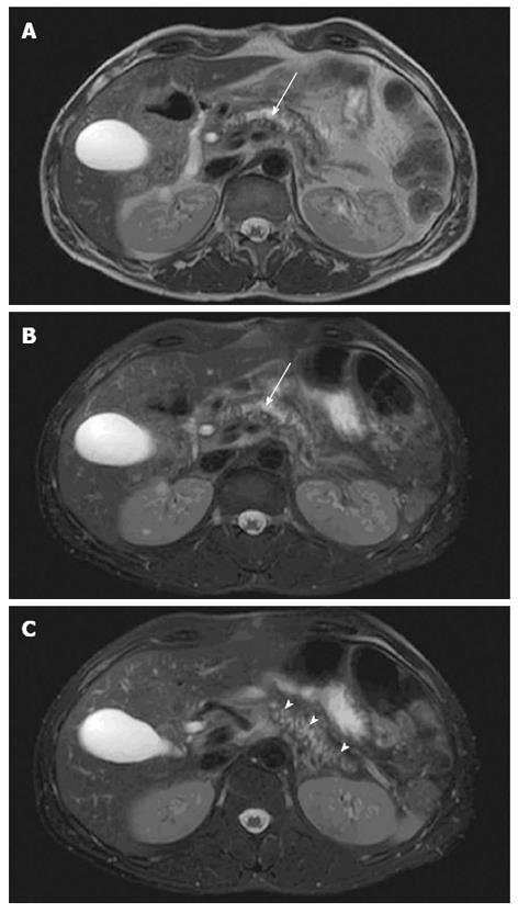
Figure 1 Pancreatic morphology.
Axial T2-weighted magnetic resonance imaging views showing glandular atrophy (A), dilated irregular duct (B, arrow) and irregular side-branches (C, arrow heads) in a patient with chronic pancreatitis.
- Citation: Hansen TM, Nilsson M, Gram M, Frøkjær JB. Morphological and functional evaluation of chronic pancreatitis with magnetic resonance imaging. World J Gastroenterol 2013; 19(42): 7241-7246
- URL: https://www.wjgnet.com/1007-9327/full/v19/i42/7241.htm
- DOI: https://dx.doi.org/10.3748/wjg.v19.i42.7241


