Copyright
©2013 Baishideng Publishing Group Co.
World J Gastroenterol. Nov 7, 2013; 19(41): 7168-7176
Published online Nov 7, 2013. doi: 10.3748/wjg.v19.i41.7168
Published online Nov 7, 2013. doi: 10.3748/wjg.v19.i41.7168
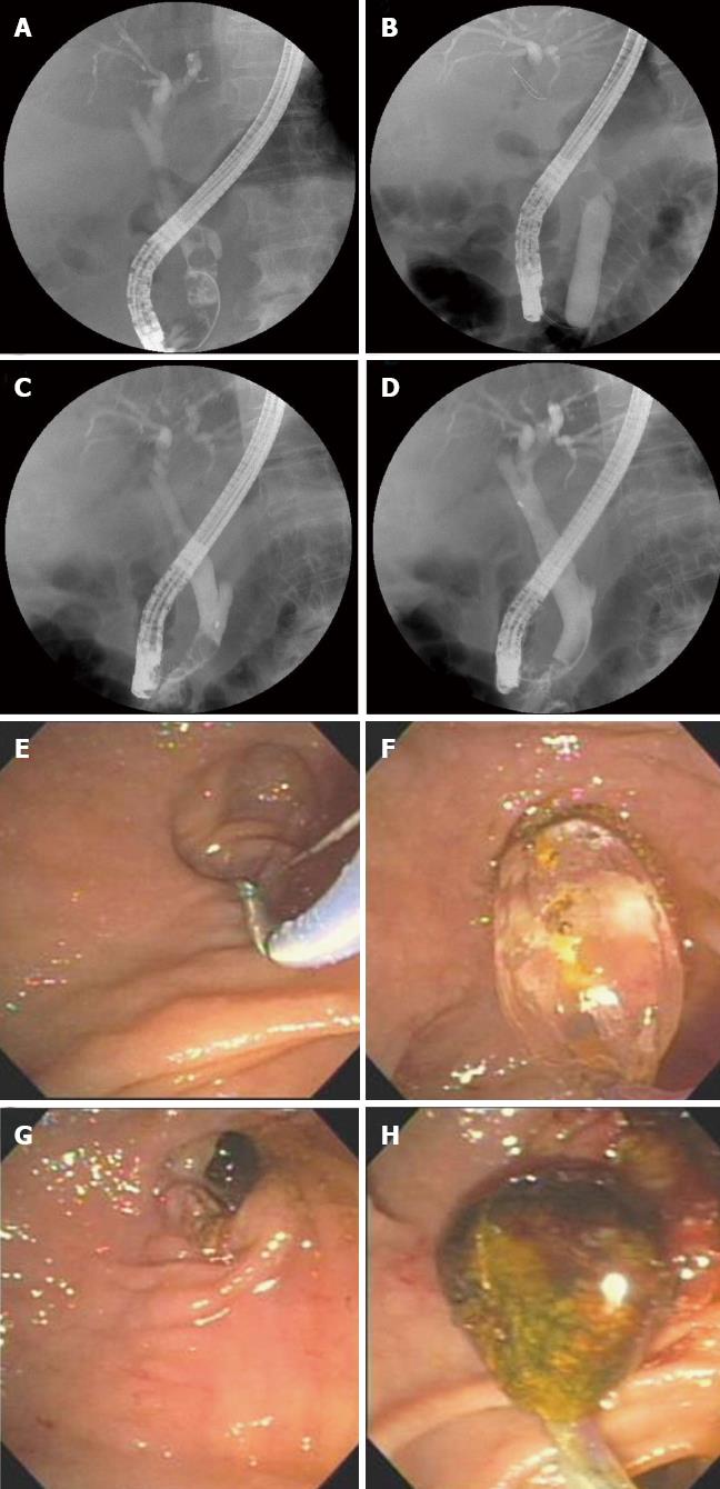
Figure 1 Endoscopic papillary large balloon dilation in a patient with periampullary diverticulum.
A: Cholangiogram showing multiple movable filling defects in the common bile duct; B: Fluoroscopic view showing the disappearance of balloon waist after gradual inflation with contrast media; C: Cholangiogram showing a large stone captured in a basket; D: Cholangiogram showing no residual filling defects in the common bile duct; E: Endoscopic sphincterotomy was carefully performed due to the presence of a huge diverticulum; F: Endoscopic view showing a dilating balloon inflated to 15 mm; G: Endoscopic view showing an enlarged ampullary opening; H: Endoscopic view showing a large brown pigment stone being extracted with a basket.
- Citation: Kim KH, Kim TN. Endoscopic papillary large balloon dilation in patients with periampullary diverticula. World J Gastroenterol 2013; 19(41): 7168-7176
- URL: https://www.wjgnet.com/1007-9327/full/v19/i41/7168.htm
- DOI: https://dx.doi.org/10.3748/wjg.v19.i41.7168


