Copyright
©2013 Baishideng Publishing Group Co.
World J Gastroenterol. Nov 7, 2013; 19(41): 7146-7153
Published online Nov 7, 2013. doi: 10.3748/wjg.v19.i41.7146
Published online Nov 7, 2013. doi: 10.3748/wjg.v19.i41.7146
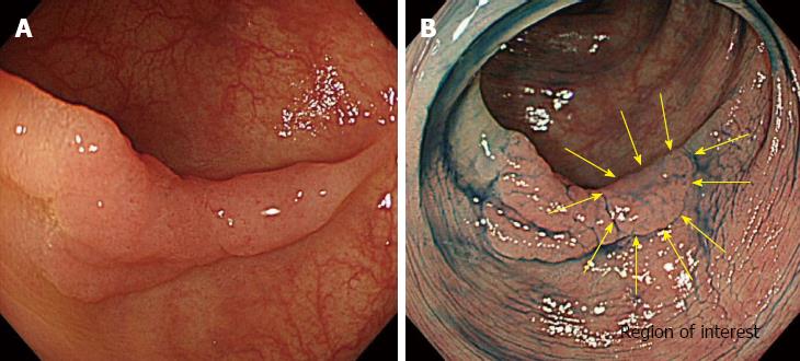
Figure 2 A determination of region of interest.
A: A white light image of a flatelevated lesion located in the sigmoid colon; B: A depressed area observed on chromoendoscopy (the area surrounded by yellow arrows). The region of interest for color tone analysis was chosen by the endoscopist based on the findings of non-magnifying narrow-band imaging or chromoendoscopy.
- Citation: Inomata H, Tamai N, Aihara H, Sumiyama K, Saito S, Kato T, Tajiri H. Efficacy of a novel auto-fluorescence imaging system with computer-assisted color analysis for assessment of colorectal lesions. World J Gastroenterol 2013; 19(41): 7146-7153
- URL: https://www.wjgnet.com/1007-9327/full/v19/i41/7146.htm
- DOI: https://dx.doi.org/10.3748/wjg.v19.i41.7146


