Copyright
©2013 Baishideng Publishing Group Co.
World J Gastroenterol. Nov 7, 2013; 19(41): 6962-6968
Published online Nov 7, 2013. doi: 10.3748/wjg.v19.i41.6962
Published online Nov 7, 2013. doi: 10.3748/wjg.v19.i41.6962
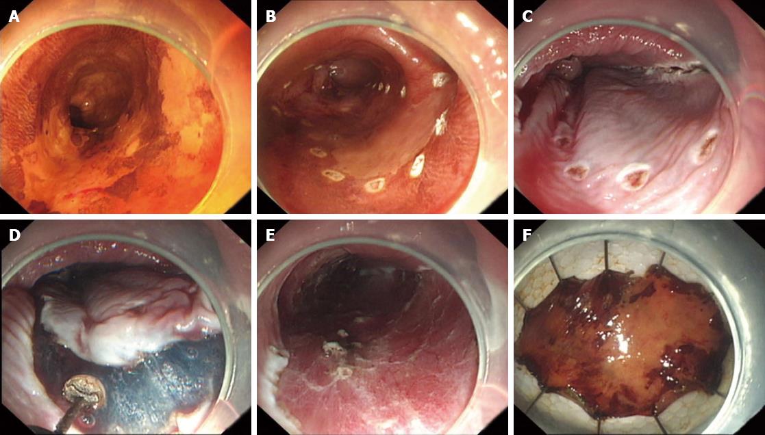
Figure 1 Process of endoscopic submucosal dissection.
A: The lesion is dyed with 2% Lugol's solution; B: Marking of the lesion by argon plasma coagulation probes; C: Submucosal injection; D: The mucosa is incised outside the marker dots, and then the submucosal tissue underneath the lesion is gradually dissected; E: The wound after resection; F: The lesion.
- Citation: Zhou PH, Shi Q, Zhong YS, Yao LQ. New progress in endoscopic treatment of esophageal diseases. World J Gastroenterol 2013; 19(41): 6962-6968
- URL: https://www.wjgnet.com/1007-9327/full/v19/i41/6962.htm
- DOI: https://dx.doi.org/10.3748/wjg.v19.i41.6962


