Copyright
©2013 Baishideng Publishing Group Co.
World J Gastroenterol. Oct 28, 2013; 19(40): 6939-6942
Published online Oct 28, 2013. doi: 10.3748/wjg.v19.i40.6939
Published online Oct 28, 2013. doi: 10.3748/wjg.v19.i40.6939
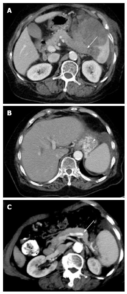
Figure 2 Imaging findings.
A: Axial contrast-enhanced abdominal computed tomography (CT) at the level of the splenic hilum shows a large low-attenuation mass in the enlarged spleen. The mass involves the splenic vein with near total occlusion (arrow); B: The development of a gastric varice (arrowhead) at the fundus; C: The 6-mo follow-up CT shows that the splenic mass has almost completely resolved, allowing the splenic vein (arrow) to return to patency.
- Citation: Chen BC, Wang HH, Lin YC, Shih YL, Chang WK, Hsieh TY. Isolated gastric variceal bleeding caused by splenic lymphoma-associated splenic vein occlusion. World J Gastroenterol 2013; 19(40): 6939-6942
- URL: https://www.wjgnet.com/1007-9327/full/v19/i40/6939.htm
- DOI: https://dx.doi.org/10.3748/wjg.v19.i40.6939


