Copyright
©2013 Baishideng Publishing Group Co.
World J Gastroenterol. Oct 28, 2013; 19(40): 6794-6804
Published online Oct 28, 2013. doi: 10.3748/wjg.v19.i40.6794
Published online Oct 28, 2013. doi: 10.3748/wjg.v19.i40.6794
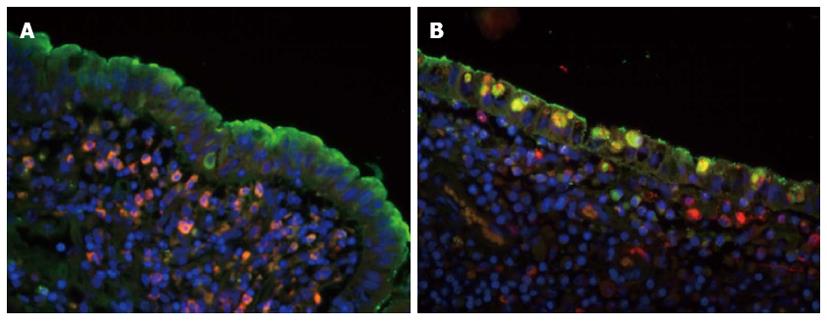
Figure 3 Co-localization of surfactant protein-A and CD68 in normal and inflammatory areas.
A: Surfactant protein (SP)-A -positive signal was indicated by fluorescence microscopy (green) fluorescence; CD68-positive signal was identified by rhodamine red-X (red) fluorescence; and the cell nucleus was indicated by Hoechst dye (blue). In the normal area, SP-A was located in the surface of the villi, specific epithelium and submucosae, lamina muscularis, mucosae and lymphoid tissues. CD68-positive cells were mainly found in the submucosae, lamina muscularis, mucosae, and lymphoid tissues and in the epithelium; B: In the inflammatory area, CD68-positive cells were dramatically increased in all levels of the bowel wall; especially CD68-positive macrophages in the epithelia of lamina mucosa. The SP-A-positive macrophages were recruited by activated epithelial cells. Double labeled SP-A and CD68 shows that some CD68-positive macrophages expressed SP-A like molecule in the inflammatory bowel disease tissues (original magnification, × 200). Figures reproduced with permission from reference [53].
- Citation: Wang H, Liu JS, Peng SH, Deng XY, Zhu DM, Javidiparsijani S, Wang GR, Li DQ, Li LX, Wang YC, Luo JM. Gut-lung crosstalk in pulmonary involvement with inflammatory bowel diseases. World J Gastroenterol 2013; 19(40): 6794-6804
- URL: https://www.wjgnet.com/1007-9327/full/v19/i40/6794.htm
- DOI: https://dx.doi.org/10.3748/wjg.v19.i40.6794


