Copyright
©2013 Baishideng Publishing Group Co.
World J Gastroenterol. Jan 28, 2013; 19(4): 597-603
Published online Jan 28, 2013. doi: 10.3748/wjg.v19.i4.597
Published online Jan 28, 2013. doi: 10.3748/wjg.v19.i4.597
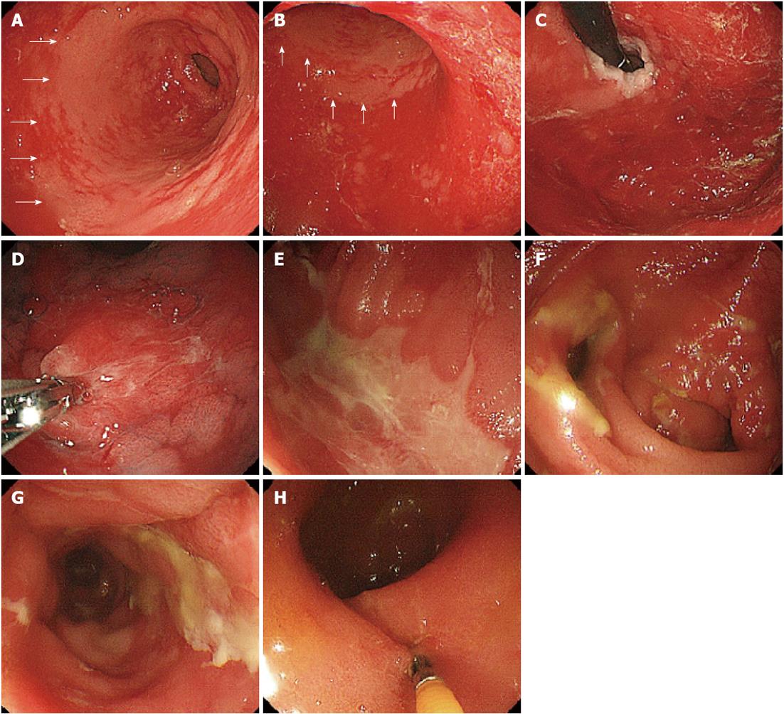
Figure 1 Upper and lower gastrointestinal endoscopic findings.
A: The gastric mucosa in the antrum of the stomach. The antrum is the only segment exhibiting normal mucosa (arrows); B: The gastric mucosa in the lower segments of the corpus of the stomach. This mucosa demonstrates diffuse sloughing, whereas the antrum is the only segment exhibiting normal mucosa (arrows); C: The gastric mucosa in the upper segments of the corpus of the stomach; D: The greater curvature of the stomach near the center. A biopsy was taken from the edematous and sloughing mucosa of the greater curvature of the upper corpus; E: Shallow ulcer with fur at the cecal valve and cecum; F: Discrete longitudinal ulcer in the terminal ileum; G: Longitudinal ulcer with fur surrounded by the inflamed edematous mucosa of the sigmoid colon; H: Edematous mucosa of the rectum.
- Citation: Okubo H, Nagata N, Uemura N. Fulminant gastrointestinal graft-versus-host disease concomitant with cytomegalovirus infection: Case report and literature review. World J Gastroenterol 2013; 19(4): 597-603
- URL: https://www.wjgnet.com/1007-9327/full/v19/i4/597.htm
- DOI: https://dx.doi.org/10.3748/wjg.v19.i4.597


