Copyright
©2013 Baishideng Publishing Group Co.
World J Gastroenterol. Jan 28, 2013; 19(4): 528-535
Published online Jan 28, 2013. doi: 10.3748/wjg.v19.i4.528
Published online Jan 28, 2013. doi: 10.3748/wjg.v19.i4.528
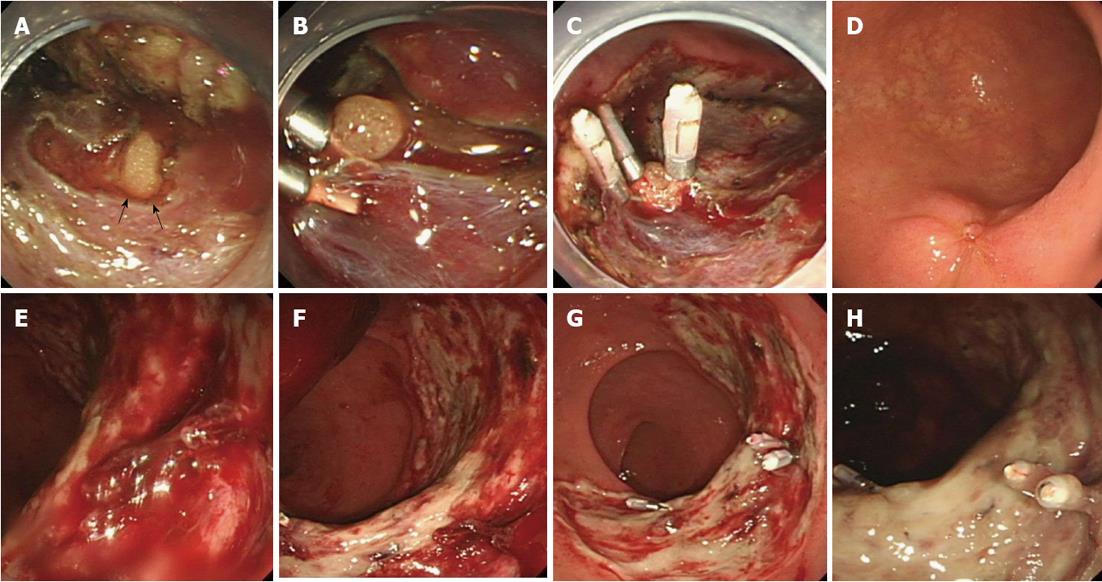
Figure 3 Representative cases of perforation and bleeding treated endoscopically.
A: A perforation was detected (arrows); B: When the omentum became visible through the perforation hole, we attempted to aspirate the omentum through the perforation hole, then clipped the gastric wall and omentum together; C: Several clips were used to seal the hole completely; D: The appearance three months after clipping; E: Bleeding from a post-endoscopic submucosal dissection ulcer was detected; F: After removing the coagulation, the bleeding point was detected; G: Clips were used to achieve hemostasis endoscopically; H: The appearance 1 wk after clipping.
- Citation: Sohara N, Hagiwara S, Arai R, Iizuka H, Onozato Y, Kakizaki S. Can endoscopic submucosal dissection be safely performed in a smaller specialized clinic? World J Gastroenterol 2013; 19(4): 528-535
- URL: https://www.wjgnet.com/1007-9327/full/v19/i4/528.htm
- DOI: https://dx.doi.org/10.3748/wjg.v19.i4.528


