Copyright
©2013 Baishideng Publishing Group Co.
World J Gastroenterol. Jan 28, 2013; 19(4): 492-502
Published online Jan 28, 2013. doi: 10.3748/wjg.v19.i4.492
Published online Jan 28, 2013. doi: 10.3748/wjg.v19.i4.492
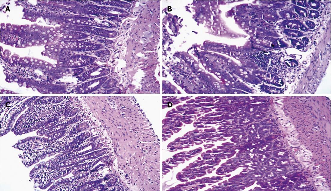
Figure 4 Hematoxylin and eosin staining of small intestine tissue.
A: Control group; B: Septic shock group; C: Early fluid resuscitation-treated group; D: Early fluid resuscitation + 2% hydrogen inhalation-treated group. Small intestinal tissues were fixed with 4% paraformaldehyde for more than 24 h. After dehydrating and embedding, they were cut into 5 μm slices, and were stained with hematoxylin and eosin. The pathogenesis of small intestine injury was observed under a light microscope (original magnification × 200). In Group A, normal intestinal mucosa was observed. In Group B, glands of small intestine were significantly damaged, and severe edema of mucosal villi, neutrophil infiltration, epithelial cell sloughing off, and even small bowel ulceration were observed. The damage mentioned above was far more severe in Group C. But in Group D, the damage was significantly reduced.
- Citation: Liu W, Shan LP, Dong XS, Liu XW, Ma T, Liu Z. Combined early fluid resuscitation and hydrogen inhalation attenuates lung and intestine injury. World J Gastroenterol 2013; 19(4): 492-502
- URL: https://www.wjgnet.com/1007-9327/full/v19/i4/492.htm
- DOI: https://dx.doi.org/10.3748/wjg.v19.i4.492


