Copyright
©2013 Baishideng Publishing Group Co.
World J Gastroenterol. Oct 21, 2013; 19(39): 6559-6567
Published online Oct 21, 2013. doi: 10.3748/wjg.v19.i39.6559
Published online Oct 21, 2013. doi: 10.3748/wjg.v19.i39.6559
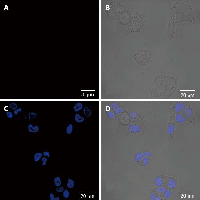
Figure 2 The fluorescence imaging of SW1990 cells with quantum dots-arginine-glycine-aspartic acid probe at the block of excess arginine-glycine-aspartic acid peptide.
A: Quantum dots- arginine-glycine-aspartic acid probe imaging; B: Original image; C: 4',6-diamidino-2-phenylindole, dihydrochloride dyeing; D: Integrated image.
- Citation: Zhou M, Ni QW, Yang SY, Qu CY, Zhao PC, Zhang JC, Xu LM. Effects of integrin-targeted photodynamic therapy on pancreatic carcinoma cell. World J Gastroenterol 2013; 19(39): 6559-6567
- URL: https://www.wjgnet.com/1007-9327/full/v19/i39/6559.htm
- DOI: https://dx.doi.org/10.3748/wjg.v19.i39.6559


