Copyright
©2013 Baishideng Publishing Group Co.
World J Gastroenterol. Oct 14, 2013; 19(38): 6505-6508
Published online Oct 14, 2013. doi: 10.3748/wjg.v19.i38.6505
Published online Oct 14, 2013. doi: 10.3748/wjg.v19.i38.6505
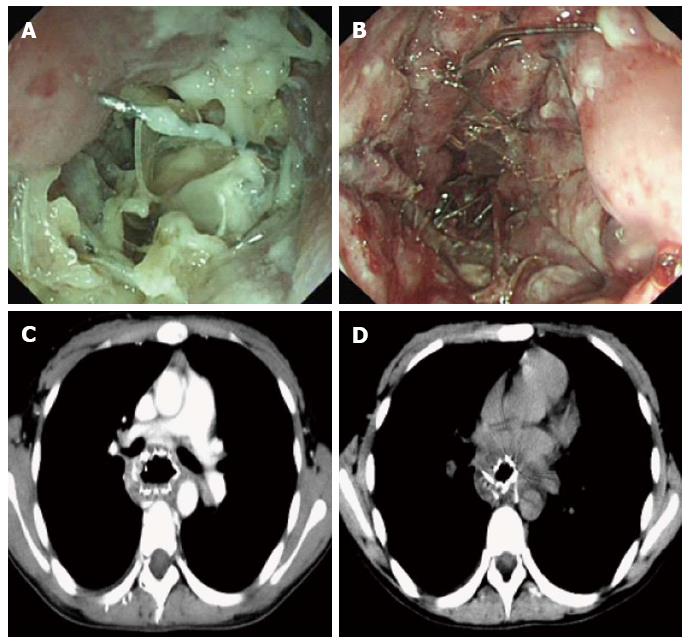
Figure 1 Disordered stent structure as a result of failure of repeated attempts at gastroscopic removal of the stent.
A-D: Enhanced chest computed tomography scan performed showed the stent was situated in the middle section of the esophagus after esophageal stent placement, the wall of the middle and inferior segments of the esophagus thickened noticeably, part of the stent was embedded into the esophageal wall, borders between the stent and surrounding fat were blurred, and the upper end of the stent pressed against the trachea carina.
- Citation: Peng GY, Kang XF, Lu X, Chen L, Zhou Q. Plastic tube-assisted gastroscopic removal of embedded esophageal metal stents: A case report. World J Gastroenterol 2013; 19(38): 6505-6508
- URL: https://www.wjgnet.com/1007-9327/full/v19/i38/6505.htm
- DOI: https://dx.doi.org/10.3748/wjg.v19.i38.6505


