Copyright
©2013 Baishideng Publishing Group Co.
World J Gastroenterol. Oct 14, 2013; 19(38): 6427-6437
Published online Oct 14, 2013. doi: 10.3748/wjg.v19.i38.6427
Published online Oct 14, 2013. doi: 10.3748/wjg.v19.i38.6427
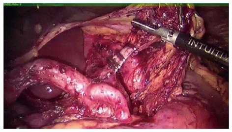
Figure 3 Lymphatic tissues are removed en bloc along the hepatic, splenic, left gastric artery and celiac trunk using an ultrasonic shear.
The origins of these arteries are clearly identified and skeletonized, and the lymphatic tissue dissected away from the adventitia. The left gastric artery is then clipped or tied at its origin.
- Citation: Liu XX, Jiang ZW, Chen P, Zhao Y, Pan HF, Li JS. Full robot-assisted gastrectomy with intracorporeal robot-sewn anastomosis produces satisfying outcomes. World J Gastroenterol 2013; 19(38): 6427-6437
- URL: https://www.wjgnet.com/1007-9327/full/v19/i38/6427.htm
- DOI: https://dx.doi.org/10.3748/wjg.v19.i38.6427


