Copyright
©2013 Baishideng Publishing Group Co.
World J Gastroenterol. Oct 7, 2013; 19(37): 6304-6309
Published online Oct 7, 2013. doi: 10.3748/wjg.v19.i37.6304
Published online Oct 7, 2013. doi: 10.3748/wjg.v19.i37.6304
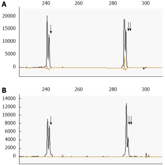
Figure 5 Polymerase chain reaction analysis from the two distinct components of the tumor demonstrated clonal immunoglobulin κ light chain gene rearrangements.
The asterisks indicate two peaks representing the rearranged polymerase chain reaction products from position 241 bp (arrow) and 281 bp (double arrows) regions of immunoglobulin κ light chain gene, respectively. A: DNA from the dissected diffuse large B-cell lymphoma component. B: DNA from the dissected classical Hodgkin lymphoma component.
- Citation: Wang HW, Yang W, Wang L, Lu YL, Lu JY. Composite diffuse large B-cell lymphoma and classical Hodgkin’s lymphoma of the stomach: Case report and literature review. World J Gastroenterol 2013; 19(37): 6304-6309
- URL: https://www.wjgnet.com/1007-9327/full/v19/i37/6304.htm
- DOI: https://dx.doi.org/10.3748/wjg.v19.i37.6304


