Copyright
©2013 Baishideng Publishing Group Co.
World J Gastroenterol. Oct 7, 2013; 19(37): 6296-6298
Published online Oct 7, 2013. doi: 10.3748/wjg.v19.i37.6296
Published online Oct 7, 2013. doi: 10.3748/wjg.v19.i37.6296
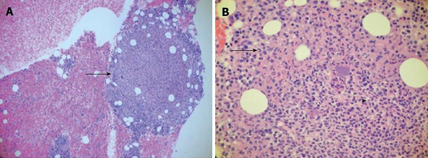
Figure 3 Bone marrow biopsy.
A: A necrotizing granuloma (arrow) with trilineage maturation and markedly increased iron storage [hematoxylin and eosin (HE) stain, ×100]; B: An area of necrosis (arrowhead) with erythrophagocytosis typical, but not diagnostic of Yersinia infection (HE stain, × 400).
- Citation: Selsky N, Forouhar F, Wu GY. An ironic case of liver infections: Yersinia enterocolitis in the setting of thalassemia. World J Gastroenterol 2013; 19(37): 6296-6298
- URL: https://www.wjgnet.com/1007-9327/full/v19/i37/6296.htm
- DOI: https://dx.doi.org/10.3748/wjg.v19.i37.6296


