Copyright
©2013 Baishideng Publishing Group Co.
World J Gastroenterol. Oct 7, 2013; 19(37): 6296-6298
Published online Oct 7, 2013. doi: 10.3748/wjg.v19.i37.6296
Published online Oct 7, 2013. doi: 10.3748/wjg.v19.i37.6296
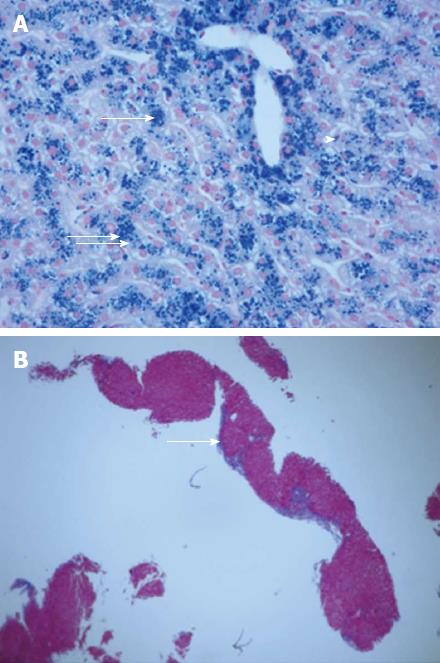
Figure 2 Liver biopsy.
A: There is marked, 3+, accumulation of iron primarily in the hepatocytes (arrows), but also in Kupffer cells (arrow head), and bile duct epithelium in association with moderate lobular hepatitis (Prussian Blue stain for iron, × 400); B: There is increased fibrosis with focal portal-to-portal and occasional central-portal septum formation (arrow) indicating progression towards early cirrhosis (Masson Trichrome stain, × 40).
- Citation: Selsky N, Forouhar F, Wu GY. An ironic case of liver infections: Yersinia enterocolitis in the setting of thalassemia. World J Gastroenterol 2013; 19(37): 6296-6298
- URL: https://www.wjgnet.com/1007-9327/full/v19/i37/6296.htm
- DOI: https://dx.doi.org/10.3748/wjg.v19.i37.6296


