Copyright
©2013 Baishideng Publishing Group Co.
World J Gastroenterol. Sep 28, 2013; 19(36): 6110-6113
Published online Sep 28, 2013. doi: 10.3748/wjg.v19.i36.6110
Published online Sep 28, 2013. doi: 10.3748/wjg.v19.i36.6110
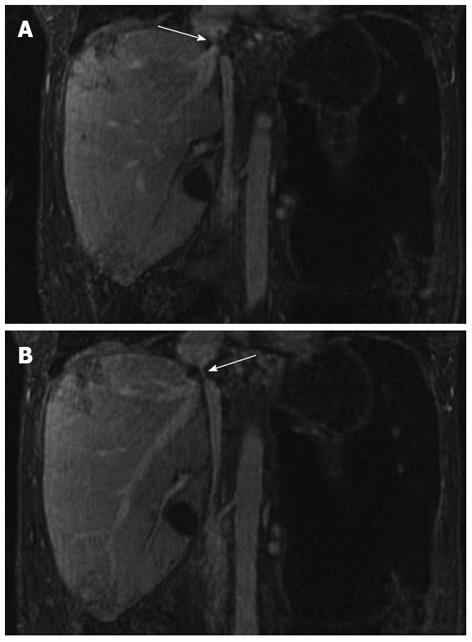
Figure 2 Five-minute steady state imaging.
A: Coronal T1 weighted ultrafast gradient echo steady state magnetic resonance imaging image obtained 5 min following administration of Gadofosveset trisodium demonstrates inferior vena cava stenosis (white arrow); B: Coronal T1 weighted ultrafast gradient echo steady state magnetic resonance imaging image obtained 5 min following administration of gadofosveset trisodium demonstrating patent, but stenosed transplant hepatic vein (white arrow).
- Citation: Strovski E, Liu D, Scudamore C, Ho S, Yoshida E, Klass D. Magnetic resonance venography and liver transplant complications. World J Gastroenterol 2013; 19(36): 6110-6113
- URL: https://www.wjgnet.com/1007-9327/full/v19/i36/6110.htm
- DOI: https://dx.doi.org/10.3748/wjg.v19.i36.6110


