Copyright
©2013 Baishideng Publishing Group Co.
World J Gastroenterol. Sep 14, 2013; 19(34): 5750-5753
Published online Sep 14, 2013. doi: 10.3748/wjg.v19.i34.5750
Published online Sep 14, 2013. doi: 10.3748/wjg.v19.i34.5750
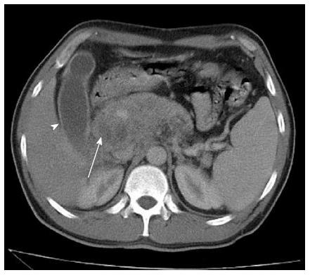
Figure 1 Contrast enhanced abdominal computed tomography.
Head, body, and tail of pancreas with enlarged dimensions and a poorly-defined margin, with two hypodense areas without significant enhancement after intravenous contrast (white arrow). The same image shows a hydropic gallbladder (arrow head) and mild splenomegaly.
- Citation: Lima TB, Domingues MAC, Caramori CA, Silva GF, Oliveira CV, Yamashiro FDS, Franzoni LC, Sassaki LY, Romeiro FG. Pancreatic paracoccidioidomycosis simulating malignant neoplasia: Case report. World J Gastroenterol 2013; 19(34): 5750-5753
- URL: https://www.wjgnet.com/1007-9327/full/v19/i34/5750.htm
- DOI: https://dx.doi.org/10.3748/wjg.v19.i34.5750


