Copyright
©2013 Baishideng Publishing Group Co.
World J Gastroenterol. Sep 7, 2013; 19(33): 5581-5585
Published online Sep 7, 2013. doi: 10.3748/wjg.v19.i33.5581
Published online Sep 7, 2013. doi: 10.3748/wjg.v19.i33.5581
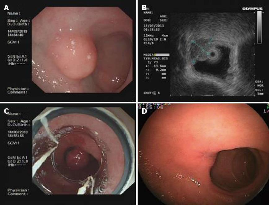
Figure 3 Imaging in case 3.
A: Endoscopic view of the protuberant lesion in the duodenal bulb; B: Endoscopic ultrasonography image of the size of the lesion; C: Endoscopic view of the ligated lesion using the endoscopic variceal ligation device; D: Endoscopic view of an ulcer scar at the ligation site after 6 wk.
- Citation: Wang L, Chen SY, Huang Y, Wu J, Leung YK. Selective endoscopic ligation for treatment of upper gastrointestinal protuberant lesions. World J Gastroenterol 2013; 19(33): 5581-5585
- URL: https://www.wjgnet.com/1007-9327/full/v19/i33/5581.htm
- DOI: https://dx.doi.org/10.3748/wjg.v19.i33.5581


