Copyright
©2013 Baishideng Publishing Group Co.
World J Gastroenterol. Aug 28, 2013; 19(32): 5385-5388
Published online Aug 28, 2013. doi: 10.3748/wjg.v19.i32.5385
Published online Aug 28, 2013. doi: 10.3748/wjg.v19.i32.5385
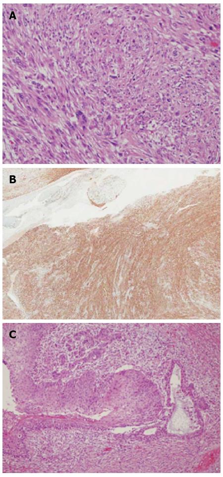
Figure 5 Pathologic images.
A: Pleomorphic spindle cells showing mitosis and cell necrosis compatible with leiomyosarcoma [hematoxylin and eosin (HE) stain, × 200]; B: Immunohistochemical stain was positive for smooth muscle actin (× 12); C: Squamous severe dysplasia and focal stratified squamous epithelial invasion into lamina propria was noted in mucosa (HE stain, × 100).
- Citation: Jang SS, Kim WT, Ko BS, Kim EH, Kim JO, Park K, Lee SW. A case of rapidly progressing leiomyosarcoma combined with squamous cell carcinoma in the esophagus. World J Gastroenterol 2013; 19(32): 5385-5388
- URL: https://www.wjgnet.com/1007-9327/full/v19/i32/5385.htm
- DOI: https://dx.doi.org/10.3748/wjg.v19.i32.5385


