Copyright
©2013 Baishideng Publishing Group Co.
World J Gastroenterol. Aug 28, 2013; 19(32): 5250-5260
Published online Aug 28, 2013. doi: 10.3748/wjg.v19.i32.5250
Published online Aug 28, 2013. doi: 10.3748/wjg.v19.i32.5250
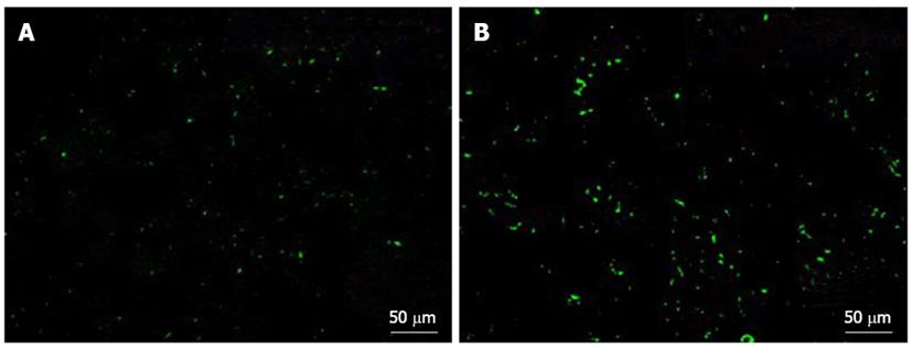
Figure 6 Accumulation of transfused human platelets in the liver.
A: Normal liver; B: Fibrotic liver. Immunostaining images obtained using anti-human CD41 antibody 2 h after transfusion. Significant human platelet accumulation in the liver was observed in the fibrotic liver, whereas few platelets accumulated in the normal liver.
- Citation: Takahashi K, Murata S, Fukunaga K, Ohkohchi N. Human platelets inhibit liver fibrosis in severe combined immunodeficiency mice. World J Gastroenterol 2013; 19(32): 5250-5260
- URL: https://www.wjgnet.com/1007-9327/full/v19/i32/5250.htm
- DOI: https://dx.doi.org/10.3748/wjg.v19.i32.5250


