Copyright
©2013 Baishideng Publishing Group Co.
World J Gastroenterol. Aug 21, 2013; 19(31): 5195-5198
Published online Aug 21, 2013. doi: 10.3748/wjg.v19.i31.5195
Published online Aug 21, 2013. doi: 10.3748/wjg.v19.i31.5195
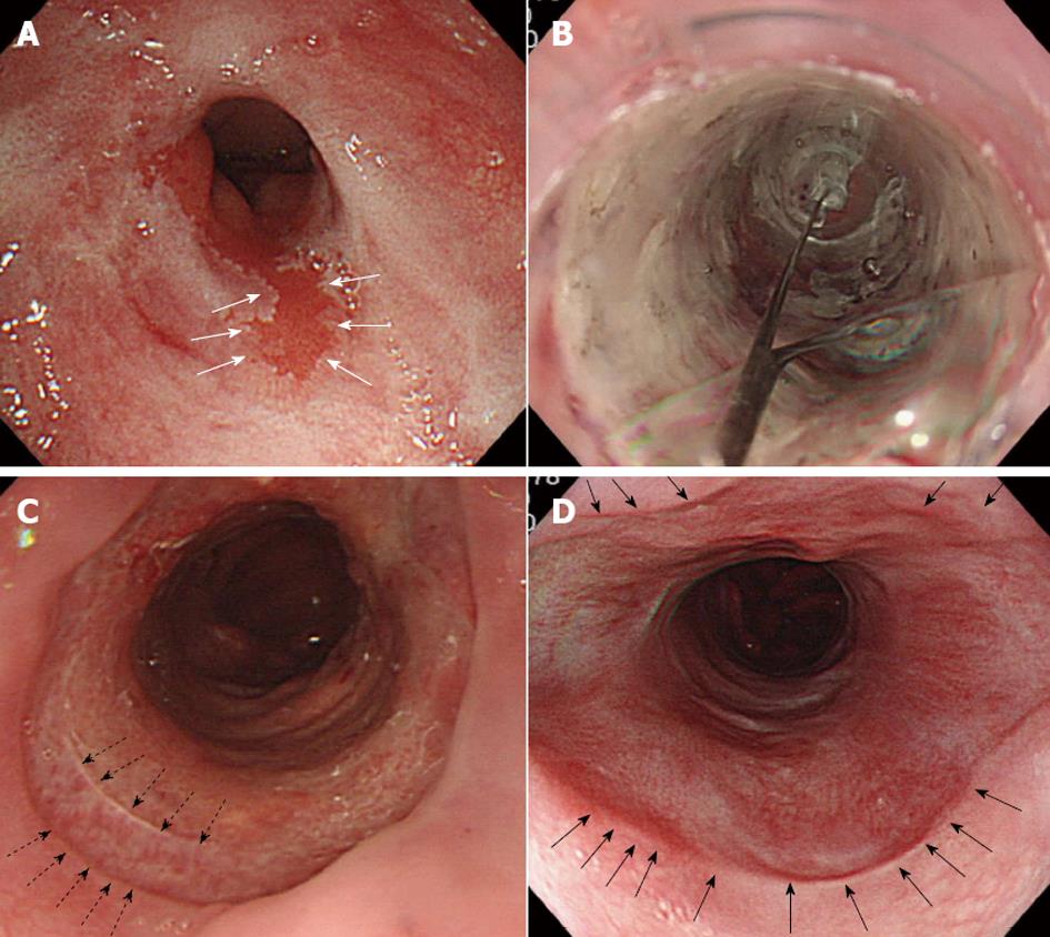
Figure 2 Barrett’s adenocarcinoma with high-grade dysplasia and severe stenosis.
A: Barrett’s adenocarcinoma (white arrows) with high-grade dysplasia and severe stenosis of the lower esophagus; B: On days 5, 8, 12, 15 and 20 after endoscopic submucosal dissection, steroid application and balloon dilatation were performed to prevent stenosis; C: The base of the artificial ulcer on day 12 after surgery. Regenerating squamous epithelium can be seen from the resection margins of the proximal side indicated by the dotted arrows; D: On day 60, the base of the artificial ulcer is thoroughly covered with regenerating squamous epithelium (black arrows). No recurrence has been observed for 5 years.
- Citation: Mori H, Kobara H, Rafiq K, Nishiyama N, Fujihara S, Ayagi M, Yachida T, Kato K, Masaki T. Radical excision of Barrett's esophagus and complete recovery of normal squamous epithelium. World J Gastroenterol 2013; 19(31): 5195-5198
- URL: https://www.wjgnet.com/1007-9327/full/v19/i31/5195.htm
- DOI: https://dx.doi.org/10.3748/wjg.v19.i31.5195


