Copyright
©2013 Baishideng Publishing Group Co.
World J Gastroenterol. Aug 21, 2013; 19(31): 5195-5198
Published online Aug 21, 2013. doi: 10.3748/wjg.v19.i31.5195
Published online Aug 21, 2013. doi: 10.3748/wjg.v19.i31.5195
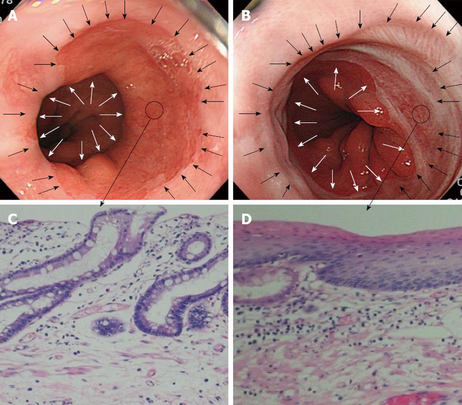
Figure 1 Short segment Barrett’s esophagus undergoing endoscopic submucosal dissection.
A: Short segment Barrett’s esophagus (SSBE) predominantly of the right sidewall. The white arrows indicate the distal side, the black arrows indicate the proximal side, and the black encircled area indicates the preoperative biopsy site; B: After circumferential resection of SSBE with endoscopic submucosal dissection: regenerating squamous epithelium is seen between the distal side (white arrows) and the proximal side (black arrows). The black encircled area indicates the postoperative biopsy site; C: Specialized columnar epithelium with the preoperative intestinal metaplasia (hematoxylin and eosin stain, × 200); D: Postoperative regenerating squamous epithelium. Specialized columnar epithelium is not seen (hematoxylin and eosin stain, × 200).
- Citation: Mori H, Kobara H, Rafiq K, Nishiyama N, Fujihara S, Ayagi M, Yachida T, Kato K, Masaki T. Radical excision of Barrett's esophagus and complete recovery of normal squamous epithelium. World J Gastroenterol 2013; 19(31): 5195-5198
- URL: https://www.wjgnet.com/1007-9327/full/v19/i31/5195.htm
- DOI: https://dx.doi.org/10.3748/wjg.v19.i31.5195


