Copyright
©2013 Baishideng Publishing Group Co.
World J Gastroenterol. Aug 14, 2013; 19(30): 4907-4916
Published online Aug 14, 2013. doi: 10.3748/wjg.v19.i30.4907
Published online Aug 14, 2013. doi: 10.3748/wjg.v19.i30.4907
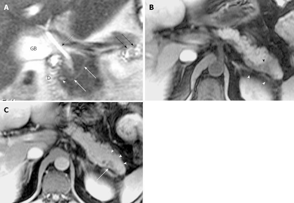
Figure 2 Magnetic resonance cholangiopancreatography and magnetic resonance imaging of recurrent acute pancreatitis with pancreas divisum only involving the pancreatic tail with a small santorinicele in a 44-year-old man with several episodes of abdominal pain.
A: Thin-section half-Fourier rapid acquisition with relaxation enhancement magnetic resonance (RARE- MR) cholangiogram [infinite/95 (effective), 3-mm section thickness] shows mild dilatation of the duct of Santorini (white arrows) with a focal enlargement consistent with santorinicele (arrowhead) at the entrance into the duodenum (D) via minor papilla. The side branch ectasia (black arrows) are noted in the pancreatic tail consistent with pancreatitis. Distended gallbladder (GB) and the CBD (dotted black arrow) are noted. B: Axial precontrast T1WI SPGR shows swelling and decrease of signal intensity in the pancreatic tail (black arrowhead), and thickening of the left anterior renal fascia (white arrowheads). C: Axial postcontrast T1WI SPGR shows mild delayed enhancement of pancreatic tail (arrowheads) with focal cystic changes (arrow).
- Citation: Wang DB, Yu J, Fulcher AS, Turner MA. Pancreatitis in patients with pancreas divisum: Imaging features at MRI and MRCP. World J Gastroenterol 2013; 19(30): 4907-4916
- URL: https://www.wjgnet.com/1007-9327/full/v19/i30/4907.htm
- DOI: https://dx.doi.org/10.3748/wjg.v19.i30.4907


