Copyright
©2013 Baishideng Publishing Group Co.
World J Gastroenterol. Aug 14, 2013; 19(30): 4897-4906
Published online Aug 14, 2013. doi: 10.3748/wjg.v19.i30.4897
Published online Aug 14, 2013. doi: 10.3748/wjg.v19.i30.4897
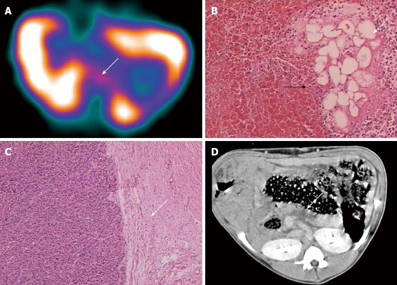
Figure 3 False-positive case of 99mTc-ciprofloxacin scintigraphy (the swine came from group B).
A: 99mTc-ciprofloxacin scintigraphy (arrow) is indicative of secondary infection (L/B = 2.15); B: However, light microscopy analysis showed pancreatic tissue and fat tissue necrosis (arrow), surrounding a marked hyperplastic area in granulated tissue (black arrow); C: Fibrous tissue (arrow), but no associated bacterial infection; D: Computed tomography image showed focal hypoattenuated areas within the pancreatic parenchyma (arrow) without gas bubbles.
- Citation: Wang JH, Sun GF, Zhang J, Shao CW, Zuo CJ, Hao J, Zheng JM, Feng XY. Infective severe acute pancreatitis: A comparison of 99mTc-ciprofloxacin scintigraphy and computed tomography. World J Gastroenterol 2013; 19(30): 4897-4906
- URL: https://www.wjgnet.com/1007-9327/full/v19/i30/4897.htm
- DOI: https://dx.doi.org/10.3748/wjg.v19.i30.4897


