Copyright
©2013 Baishideng Publishing Group Co.
World J Gastroenterol. Aug 14, 2013; 19(30): 4850-4860
Published online Aug 14, 2013. doi: 10.3748/wjg.v19.i30.4850
Published online Aug 14, 2013. doi: 10.3748/wjg.v19.i30.4850
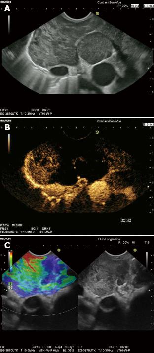
Figure 8 Endosonography.
Enlarged subcarinal lymph nodes in Non Hodgkin Lymphoma (A: B-Mode). Contrast enhanced endoscopic ultrasound demonstrates extensive avascular (necrotic) areas (B) and real time endoscopic indicates hard (blue), infiltrated areas (C), thus targeting endoscopic ultrasound-guided biopsy.
- Citation: Cui XW, Jenssen C, Saftoiu A, Ignee A, Dietrich CF. New ultrasound techniques for lymph node evaluation. World J Gastroenterol 2013; 19(30): 4850-4860
- URL: https://www.wjgnet.com/1007-9327/full/v19/i30/4850.htm
- DOI: https://dx.doi.org/10.3748/wjg.v19.i30.4850


