Copyright
©2013 Baishideng Publishing Group Co.
World J Gastroenterol. Aug 14, 2013; 19(30): 4850-4860
Published online Aug 14, 2013. doi: 10.3748/wjg.v19.i30.4850
Published online Aug 14, 2013. doi: 10.3748/wjg.v19.i30.4850
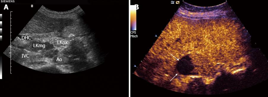
Figure 1 Lymph node infiltration, carcinoma.
A: With lymph node (LN) specific contrast agents malignant infiltration can be delineated (LKmg) as focal hypoenhancement in the upper part of this perihepatic LN. The lower part (LKnx) shows normal (physiological) enhancement; B: With SonoVue®. Necrotic (non-enhancing, arrows) areas can be detected within this perihepatic lymph node. Necrotic areas are typically for carcinoma infiltration and tuberculosis. IVC: Inferior vena cava; Ao: Aorta; DHC: Common bile duct.
- Citation: Cui XW, Jenssen C, Saftoiu A, Ignee A, Dietrich CF. New ultrasound techniques for lymph node evaluation. World J Gastroenterol 2013; 19(30): 4850-4860
- URL: https://www.wjgnet.com/1007-9327/full/v19/i30/4850.htm
- DOI: https://dx.doi.org/10.3748/wjg.v19.i30.4850


