Copyright
©2013 Baishideng Publishing Group Co.
World J Gastroenterol. Jan 21, 2013; 19(3): 422-425
Published online Jan 21, 2013. doi: 10.3748/wjg.v19.i3.422
Published online Jan 21, 2013. doi: 10.3748/wjg.v19.i3.422
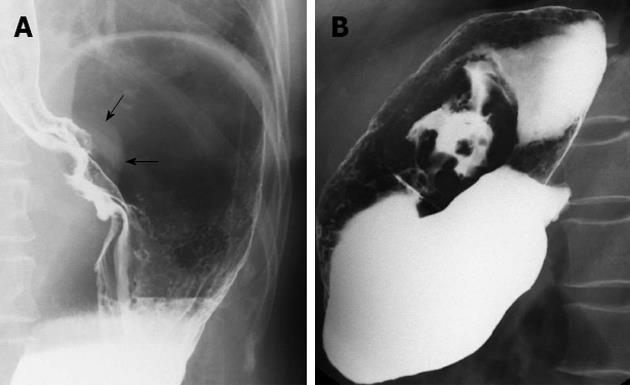
Figure 3 Double contrast examination demonstrated the shadow of soft tissue mass around the cardia and lesser curvature of stomach (arrows) (A), and filling defect, intracavitary niches, destruction of mucosa, as well as rigidity of the local wall were seen (B).
The abdominal part of esophagus was involved.
- Citation: Shen XZ, Liu F, Ni RJ, Wang BY. Primary histiocytic sarcoma of the stomach: A case report with imaging findings. World J Gastroenterol 2013; 19(3): 422-425
- URL: https://www.wjgnet.com/1007-9327/full/v19/i3/422.htm
- DOI: https://dx.doi.org/10.3748/wjg.v19.i3.422


