Copyright
©2013 Baishideng Publishing Group Co.
World J Gastroenterol. Aug 7, 2013; 19(29): 4836-4840
Published online Aug 7, 2013. doi: 10.3748/wjg.v19.i29.4836
Published online Aug 7, 2013. doi: 10.3748/wjg.v19.i29.4836
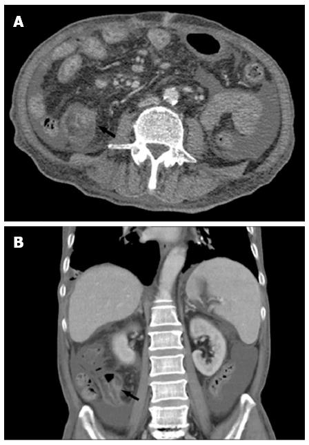
Figure 3 Abdominal computed tomography showed mild intestinal dilatation and wall thickening in the terminal ileum (marked with black arrow), and moderate ascites in the abdominal cavity.
A: Horizontal slice; B: Coronal slice.
- Citation: Gao X, Wang ZJ. Idiopathic chronic ulcerative enteritis with perforation and recurrent bleeding: A case report. World J Gastroenterol 2013; 19(29): 4836-4840
- URL: https://www.wjgnet.com/1007-9327/full/v19/i29/4836.htm
- DOI: https://dx.doi.org/10.3748/wjg.v19.i29.4836


