Copyright
©2013 Baishideng Publishing Group Co.
World J Gastroenterol. Aug 7, 2013; 19(29): 4774-4780
Published online Aug 7, 2013. doi: 10.3748/wjg.v19.i29.4774
Published online Aug 7, 2013. doi: 10.3748/wjg.v19.i29.4774
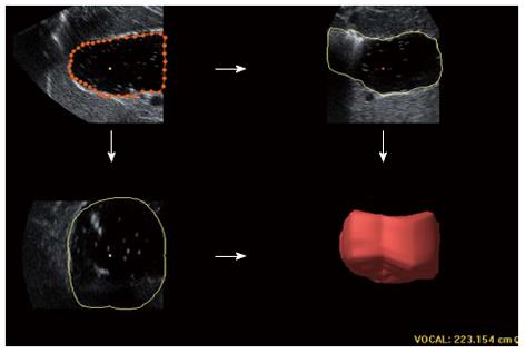
Figure 3 Three-dimensional ultrasound applied for measuring proximal gastric volume.
The volume is measured similarly from the top inner margin of the fundus to 7 cm level inferiorly along the long axis of proximal stomach; six sections of the block from six 30° rotations are separately outlined along the echoic interface in the upper left view. A reconstructive volume is displayed in the lower right view.
- Citation: Fan XP, Wang L, Zhu Q, Ma T, Xia CX, Zhou YJ. Sonographic evaluation of proximal gastric accommodation in patients with functional dyspepsia. World J Gastroenterol 2013; 19(29): 4774-4780
- URL: https://www.wjgnet.com/1007-9327/full/v19/i29/4774.htm
- DOI: https://dx.doi.org/10.3748/wjg.v19.i29.4774


