Copyright
©2013 Baishideng Publishing Group Co.
World J Gastroenterol. Aug 7, 2013; 19(29): 4774-4780
Published online Aug 7, 2013. doi: 10.3748/wjg.v19.i29.4774
Published online Aug 7, 2013. doi: 10.3748/wjg.v19.i29.4774
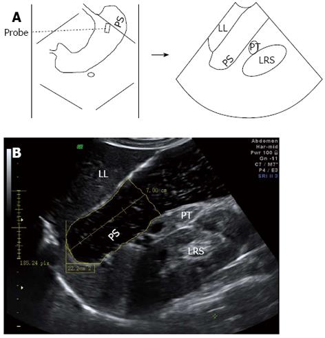
Figure 1 Sagittal section of the proximal stomach.
A: To obtain the section, a probe is placed longitudinally under the left subcostal margin and tilted cranially in the long axial direction of proximal stomach (PS) to show the top of gastric fundus, in which left renal sinus (LRS), left liver (LL), and pancreatic tail (PT) are simultaneously displayed; B: Proximal gastric area (PGA) is measured by means of outlining along the echogenic mucosa surface of PS in the distance between the echoic inner surface of the fundus top down to 7 cm level (between cursors).
- Citation: Fan XP, Wang L, Zhu Q, Ma T, Xia CX, Zhou YJ. Sonographic evaluation of proximal gastric accommodation in patients with functional dyspepsia. World J Gastroenterol 2013; 19(29): 4774-4780
- URL: https://www.wjgnet.com/1007-9327/full/v19/i29/4774.htm
- DOI: https://dx.doi.org/10.3748/wjg.v19.i29.4774


