Copyright
©2013 Baishideng Publishing Group Co.
World J Gastroenterol. Jul 28, 2013; 19(28): 4624-4629
Published online Jul 28, 2013. doi: 10.3748/wjg.v19.i28.4624
Published online Jul 28, 2013. doi: 10.3748/wjg.v19.i28.4624
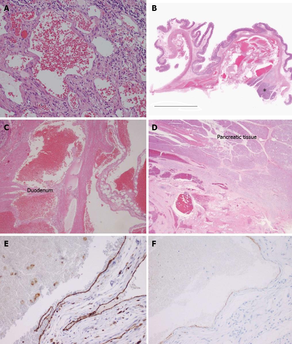
Figure 4 Pathological analysis by hematoxylin and eosin staining and immunohistochemistry.
A: A representative tissue section from the main region of the cavernous hemangioma [hematoxylin and eosin (HE), magnification × 200]; B: The loupe view demonstrates a portion of cavernous hemangioma infiltrating the pancreatic head (asterisk) and duodenal wall (HE). The scale represents 10 mm; C: Tumor invasion into the muscle layer of the duodenum (HE, magnification × 4); D: Tumor invasion into the pancreas head (HE, magnification × 1); E: Positive immunostaining for CD31 supports the diagnosis of hemangioma (magnification × 20); F: The lumen showed partial and weakly positive staining for podoplanin/D2-40 (magnification × 20).
- Citation: Hanaoka M, Hashimoto M, Sasaki K, Matsuda M, Fujii T, Ohashi K, Watanabe G. Retroperitoneal cavernous hemangioma resected by a pylorus preserving pancreaticoduodenectomy. World J Gastroenterol 2013; 19(28): 4624-4629
- URL: https://www.wjgnet.com/1007-9327/full/v19/i28/4624.htm
- DOI: https://dx.doi.org/10.3748/wjg.v19.i28.4624


