Copyright
©2013 Baishideng Publishing Group Co.
World J Gastroenterol. Jul 28, 2013; 19(28): 4624-4629
Published online Jul 28, 2013. doi: 10.3748/wjg.v19.i28.4624
Published online Jul 28, 2013. doi: 10.3748/wjg.v19.i28.4624
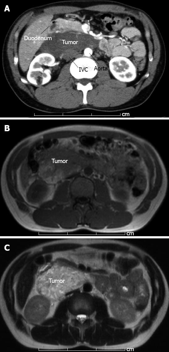
Figure 1 12 cm × 9 cm tumor was detected by computed tomography and magnetic resonance imaging.
A: Abdominal enhanced computed tomography in early phase showed tumor without marked contrast. The tumor had pushed the pancreas to the ventral side; B: T1-weighted image of magnetic resonance imaging showed low and relatively high intensity area inside the tumor; C: T2-weighted image showed high intensity area with a few part of intermediate signal intensity area. IVC: Inferior vena cava.
- Citation: Hanaoka M, Hashimoto M, Sasaki K, Matsuda M, Fujii T, Ohashi K, Watanabe G. Retroperitoneal cavernous hemangioma resected by a pylorus preserving pancreaticoduodenectomy. World J Gastroenterol 2013; 19(28): 4624-4629
- URL: https://www.wjgnet.com/1007-9327/full/v19/i28/4624.htm
- DOI: https://dx.doi.org/10.3748/wjg.v19.i28.4624


