Copyright
©2013 Baishideng Publishing Group Co.
World J Gastroenterol. Jul 28, 2013; 19(28): 4616-4623
Published online Jul 28, 2013. doi: 10.3748/wjg.v19.i28.4616
Published online Jul 28, 2013. doi: 10.3748/wjg.v19.i28.4616
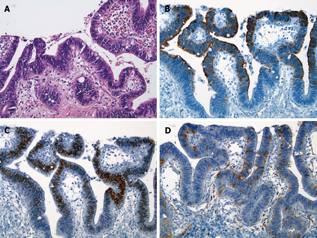
Figure 3 Histologic features of the dysplastic epithelium found in a medium-sized perihilar bile duct located within the invasive neoplasm.
A: The dysplastic epithelium contained some goblet cells [Hematoxylin and eosin (HE), × 200]; B, C: The dysplastic epithelial cells were immunoreactive for CK20 (B, × 200) and CDX2 (C, × 200); D: Some of the dysplastic epithelial cells were positive for synaptophysin with less extensive and weak immunoreactivity compared to the invasive neoplasm (× 200).
- Citation: Sasatomi E, Nalesnik MA, Marsh JW. Neuroendocrine carcinoma of the extrahepatic bile duct: Case report and literature review. World J Gastroenterol 2013; 19(28): 4616-4623
- URL: https://www.wjgnet.com/1007-9327/full/v19/i28/4616.htm
- DOI: https://dx.doi.org/10.3748/wjg.v19.i28.4616


