Copyright
©2013 Baishideng Publishing Group Co.
World J Gastroenterol. Jul 28, 2013; 19(28): 4616-4623
Published online Jul 28, 2013. doi: 10.3748/wjg.v19.i28.4616
Published online Jul 28, 2013. doi: 10.3748/wjg.v19.i28.4616
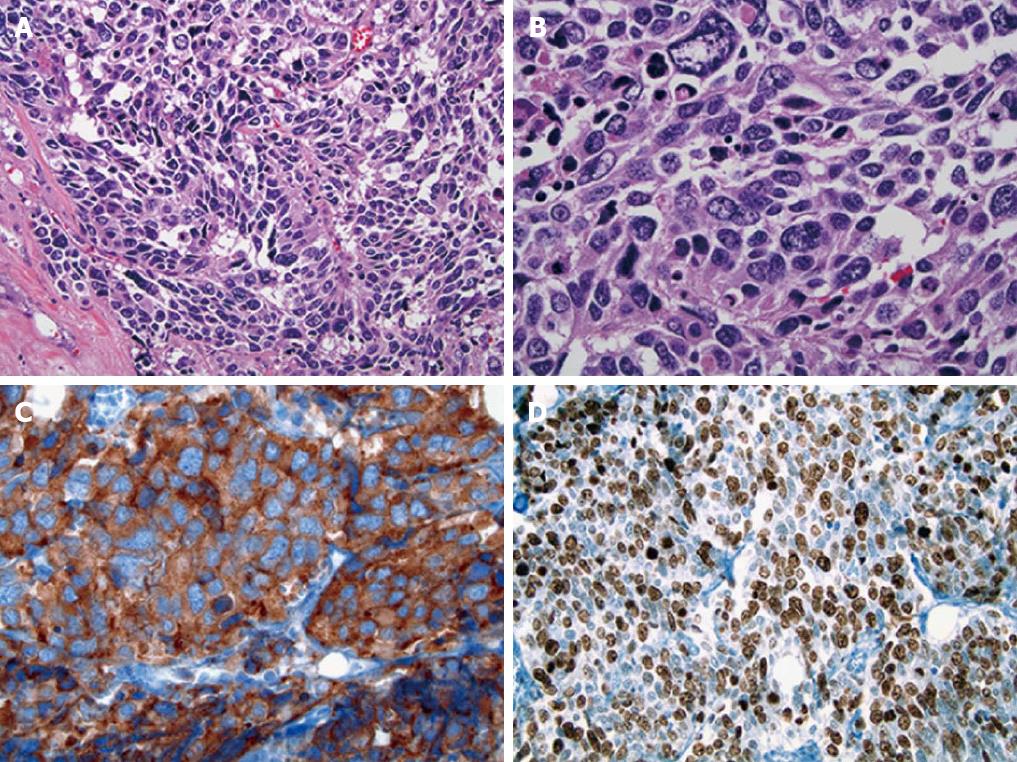
Figure 2 Histologic features of the hilar neoplasm.
A: The tumor cells were arranged in cellular nests and sheets with little intervening fibrovascular stroma [Hematoxylin and eosin (HE), × 200]; B: The tumor cells had a small to moderate amount of amphophilic cytoplasm and medium to large hyperchromatic nuclei with fine to coarse granular chromatin and occasional small nucleoli (HE, × 400); C: The tumor cells were strongly positive for synaptophysin (× 400); D: 70%-80% of the tumor cells were positive for Ki-67 (× 200).
- Citation: Sasatomi E, Nalesnik MA, Marsh JW. Neuroendocrine carcinoma of the extrahepatic bile duct: Case report and literature review. World J Gastroenterol 2013; 19(28): 4616-4623
- URL: https://www.wjgnet.com/1007-9327/full/v19/i28/4616.htm
- DOI: https://dx.doi.org/10.3748/wjg.v19.i28.4616


