Copyright
©2013 Baishideng Publishing Group Co.
World J Gastroenterol. Jul 28, 2013; 19(28): 4475-4485
Published online Jul 28, 2013. doi: 10.3748/wjg.v19.i28.4475
Published online Jul 28, 2013. doi: 10.3748/wjg.v19.i28.4475
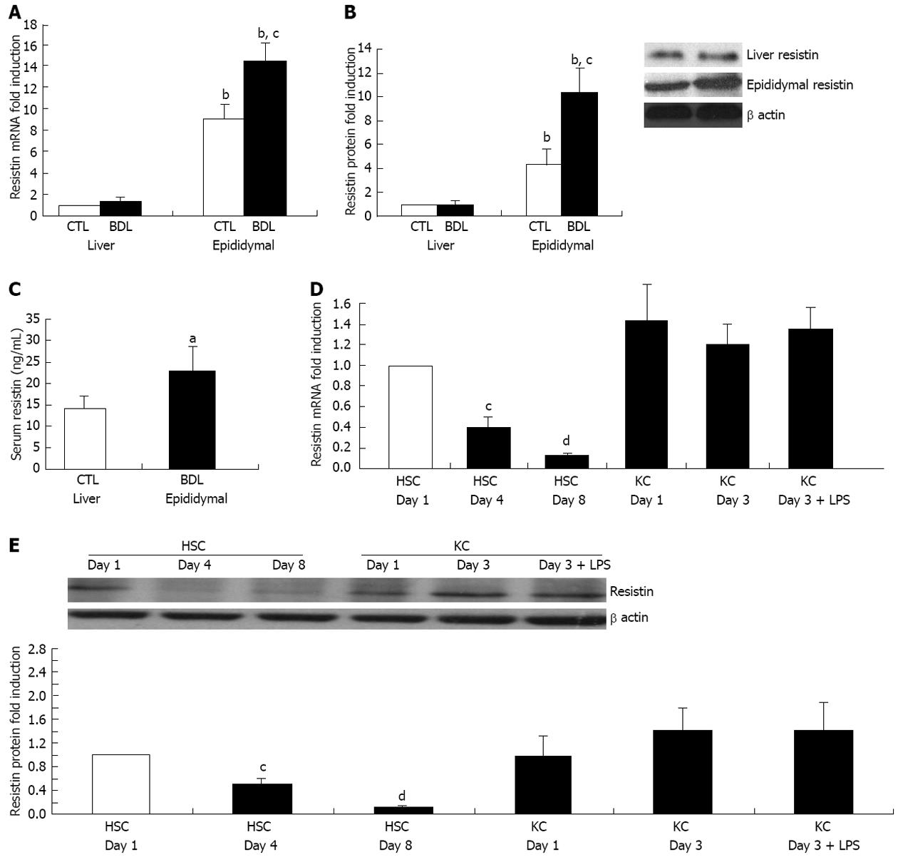
Figure 1 Rat extrahepatic but not intrahepatic resistin is up-regulated in cirrhosis.
For the in vivo animal study, Sprague Dawley rats were subjected to bile duct ligation (BDL) for 4 wk. Liver, adipose tissue (epididymal fat) and serum were collected to determine resistin expression by quantitative polymerase chain reaction (qPCR), immunoblot and enzyme-linked immunosorbent assay. For the cell culture study, rat hepatic stellate cells (HSCs) and Kupffer cells (KCs) were isolated and cultured on plastic. HSC and KC total RNA/protein were extracted at different culture times (day 1, 4 and 8 for HSCs; day 1 and 3 for KCs). One group of KCs at day 2 were treated with Lipopolysaccharide (LPS) (50 ng/mL) for 24 h. qPCR and Immunoblot were performed for quantification of resistin mRNA and protein. β actin was used as an internal control. A: mRNA expression of resistin in liver and epididymal fat; B: Protein expression of resistin in liver and epididymal fat; C: Serum resistin concentrations; D: mRNA expression of resistin in HSCs and KCs on different culture days; E: Protein expression of resistin in HSCs and KCs on different culture days. Results are mean ± SD of at least three independent experiments performed in triplicate. aP < 0.05 and bP < 0.01 increased vs rat liver, HSC control at day 1 or sham rat serum; cP < 0.05 and dP < 0.01 decreased vs liver of control sham rat or HSC control at day 1.
- Citation: Dong ZX, Su L, Brymora J, Bird C, Xie Q, George J, Wang JH. Resistin mediates the hepatic stellate cell phenotype. World J Gastroenterol 2013; 19(28): 4475-4485
- URL: https://www.wjgnet.com/1007-9327/full/v19/i28/4475.htm
- DOI: https://dx.doi.org/10.3748/wjg.v19.i28.4475


