Copyright
©2013 Baishideng Publishing Group Co.
World J Gastroenterol. Jul 21, 2013; 19(27): 4437-4442
Published online Jul 21, 2013. doi: 10.3748/wjg.v19.i27.4437
Published online Jul 21, 2013. doi: 10.3748/wjg.v19.i27.4437
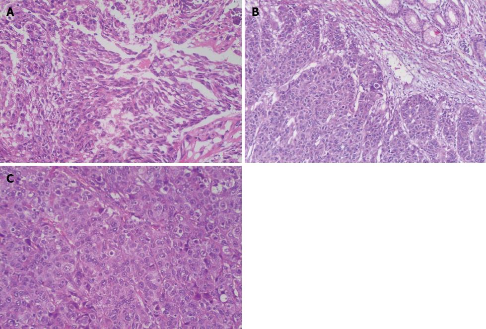
Figure 3 Presentations of hematoxylin and eosin stains: A: Tumor cells are arranged in a trabecular pattern (× 200); B: Tumor cells are arranged with cancer nests and adenoids (× 100); C: Tumor cells are featured with eosinophilic cytoplasm and round nuclei occasionally exhibiting obvious nucleoli (× 200).
- Citation: Ye MF, Tao F, Liu F, Sun AJ. Hepatoid adenocarcinoma of the stomach: A report of three cases. World J Gastroenterol 2013; 19(27): 4437-4442
- URL: https://www.wjgnet.com/1007-9327/full/v19/i27/4437.htm
- DOI: https://dx.doi.org/10.3748/wjg.v19.i27.4437


