Copyright
©2013 Baishideng Publishing Group Co.
World J Gastroenterol. Jul 21, 2013; 19(27): 4316-4324
Published online Jul 21, 2013. doi: 10.3748/wjg.v19.i27.4316
Published online Jul 21, 2013. doi: 10.3748/wjg.v19.i27.4316
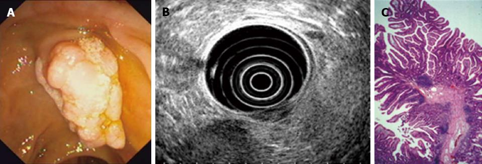
Figure 2 Diagnostic workup of a representative case (tubulovillous adenoma of the papilla).
A: Initial endoscopic view onto the tumor-like lesion of papillary region; B: Endoscopic ultrasonography-based imaging indicating echo-poor tumor lesion but no infiltrating tumor growth; C: Histopathological picture (HE staining; magnification, × 40).
- Citation: Will U, Müller AK, Fueldner F, Wanzar I, Meyer F. Endoscopic papillectomy: Data of a prospective observational study. World J Gastroenterol 2013; 19(27): 4316-4324
- URL: https://www.wjgnet.com/1007-9327/full/v19/i27/4316.htm
- DOI: https://dx.doi.org/10.3748/wjg.v19.i27.4316


