Copyright
©2013 Baishideng Publishing Group Co.
World J Gastroenterol. Jul 21, 2013; 19(27): 4316-4324
Published online Jul 21, 2013. doi: 10.3748/wjg.v19.i27.4316
Published online Jul 21, 2013. doi: 10.3748/wjg.v19.i27.4316
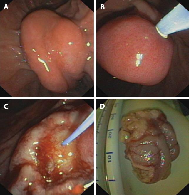
Figure 1 Steps of endoscopic papillectomy.
A: Endoscopic view onto the local tumor site (adenomatous papilla of Vater); B: Insertion of a tube through the papilla of Vater for cholangiography; C: Postinterventional endoscopic view onto the papillary region after endoscopic papillectomy; D: Tumor specimen ex situ.
- Citation: Will U, Müller AK, Fueldner F, Wanzar I, Meyer F. Endoscopic papillectomy: Data of a prospective observational study. World J Gastroenterol 2013; 19(27): 4316-4324
- URL: https://www.wjgnet.com/1007-9327/full/v19/i27/4316.htm
- DOI: https://dx.doi.org/10.3748/wjg.v19.i27.4316


