Copyright
©2013 Baishideng Publishing Group Co.
World J Gastroenterol. Jul 21, 2013; 19(27): 4300-4308
Published online Jul 21, 2013. doi: 10.3748/wjg.v19.i27.4300
Published online Jul 21, 2013. doi: 10.3748/wjg.v19.i27.4300
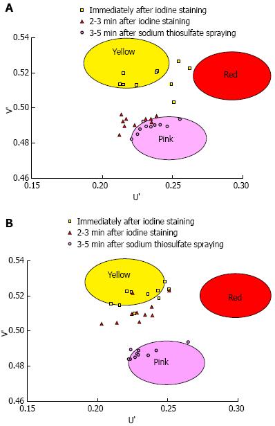
Figure 4 The U’ and V’ values of the pink-color sign positive mucosa and negative mucosa in the early, late and final phases were plotted on a color diagram.
A: Pink-color sign positive mucosa; B: Pink-color sign negative mucosa.
- Citation: Ishihara R, Kanzaki H, Iishi H, Nagai K, Matsui F, Yamashina T, Matsuura N, Ito T, Fujii M, Yamamoto S, Hanaoka N, Takeuchi Y, Higashino K, Uedo N, Tatsuta M, Tomita Y, Ishiguro S. Pink-color sign in esophageal squamous neoplasia, and speculation regarding the underlying mechanism. World J Gastroenterol 2013; 19(27): 4300-4308
- URL: https://www.wjgnet.com/1007-9327/full/v19/i27/4300.htm
- DOI: https://dx.doi.org/10.3748/wjg.v19.i27.4300


