Copyright
©2013 Baishideng Publishing Group Co.
World J Gastroenterol. Jul 21, 2013; 19(27): 4300-4308
Published online Jul 21, 2013. doi: 10.3748/wjg.v19.i27.4300
Published online Jul 21, 2013. doi: 10.3748/wjg.v19.i27.4300
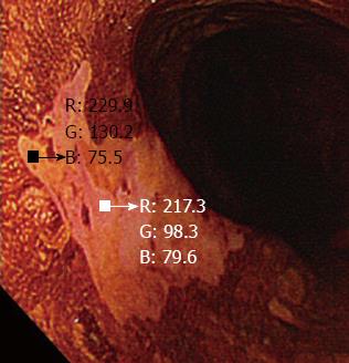
Figure 3 A small region of interest was chosen in both the pink-color sign positive and pink-color sign negative areas.
These regions of interest were carefully chosen to be of similar size in each area. The red-green-blue components of each region of interest were calculated using Image J software.
- Citation: Ishihara R, Kanzaki H, Iishi H, Nagai K, Matsui F, Yamashina T, Matsuura N, Ito T, Fujii M, Yamamoto S, Hanaoka N, Takeuchi Y, Higashino K, Uedo N, Tatsuta M, Tomita Y, Ishiguro S. Pink-color sign in esophageal squamous neoplasia, and speculation regarding the underlying mechanism. World J Gastroenterol 2013; 19(27): 4300-4308
- URL: https://www.wjgnet.com/1007-9327/full/v19/i27/4300.htm
- DOI: https://dx.doi.org/10.3748/wjg.v19.i27.4300


