Copyright
©2013 Baishideng Publishing Group Co.
World J Gastroenterol. Jul 21, 2013; 19(27): 4300-4308
Published online Jul 21, 2013. doi: 10.3748/wjg.v19.i27.4300
Published online Jul 21, 2013. doi: 10.3748/wjg.v19.i27.4300
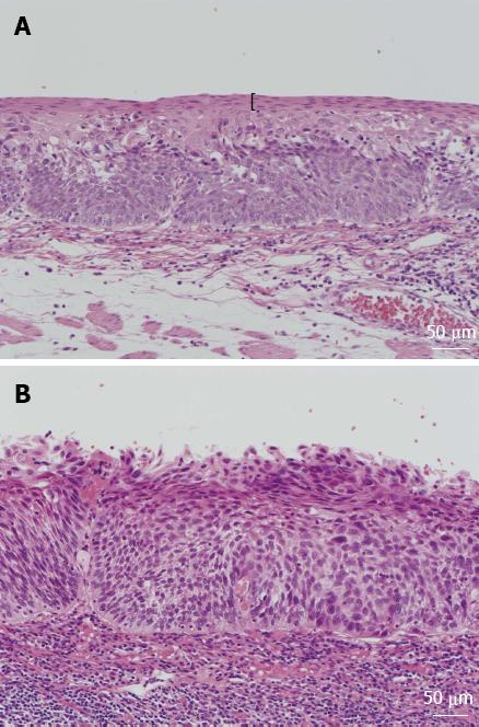
Figure 1 Histologic findings of esophageal lesions.
A: Esophageal lesion with keratinous layers, shown by black parentheses; B: Esophageal lesion without keratinous layers.
- Citation: Ishihara R, Kanzaki H, Iishi H, Nagai K, Matsui F, Yamashina T, Matsuura N, Ito T, Fujii M, Yamamoto S, Hanaoka N, Takeuchi Y, Higashino K, Uedo N, Tatsuta M, Tomita Y, Ishiguro S. Pink-color sign in esophageal squamous neoplasia, and speculation regarding the underlying mechanism. World J Gastroenterol 2013; 19(27): 4300-4308
- URL: https://www.wjgnet.com/1007-9327/full/v19/i27/4300.htm
- DOI: https://dx.doi.org/10.3748/wjg.v19.i27.4300


