Copyright
©2013 Baishideng Publishing Group Co.
World J Gastroenterol. Jul 14, 2013; 19(26): 4192-4199
Published online Jul 14, 2013. doi: 10.3748/wjg.v19.i26.4192
Published online Jul 14, 2013. doi: 10.3748/wjg.v19.i26.4192
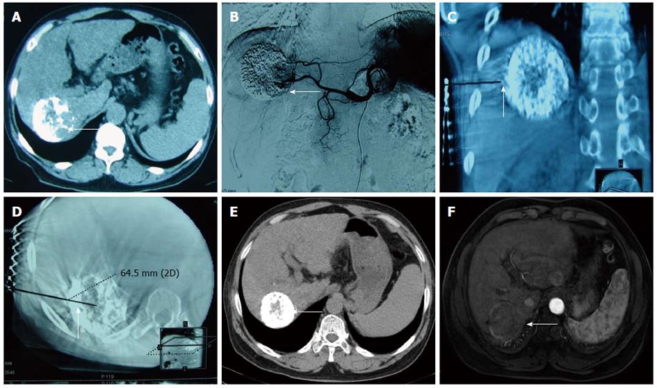
Figure 1 A male patient aged 60 years with hepatocellular carcinoma.
Computed tomography (CT) scan showed a residual lesion in the liver after the first interventional therapy. Thus, transcatheter arterial chemoembolisation (TACE) immediately followed by radiofrequency ablation (RFA) was performed under the guidance of digital subtraction angiography (DSA)-CT. A: Liver CT scan showed a partial lipiodol deposit (arrow) after first interventional therapy; B: Angiogram performed before RFA showed a lipiodol deposit in the liver and staining of the delay phase around the lipiodol (arrow); C, D: INNOVA4100 IQ DSA-CT was used to obtain the coronal section and cross section of the reconstructed image to design the puncture path and angle (arrow); E, F: Liver CT scan 23 mo after combination therapy showed good lipiodol deposits without enhancement (arrow).
- Citation: Wang ZJ, Wang MQ, Duan F, Song P, Liu FY, Chang ZF, Wang Y, Yan JY, Li K. Transcatheter arterial chemoembolization followed by immediate radiofrequency ablation for large solitary hepatocellular carcinomas. World J Gastroenterol 2013; 19(26): 4192-4199
- URL: https://www.wjgnet.com/1007-9327/full/v19/i26/4192.htm
- DOI: https://dx.doi.org/10.3748/wjg.v19.i26.4192


