Copyright
©2013 Baishideng Publishing Group Co.
World J Gastroenterol. Jul 14, 2013; 19(26): 4127-4136
Published online Jul 14, 2013. doi: 10.3748/wjg.v19.i26.4127
Published online Jul 14, 2013. doi: 10.3748/wjg.v19.i26.4127
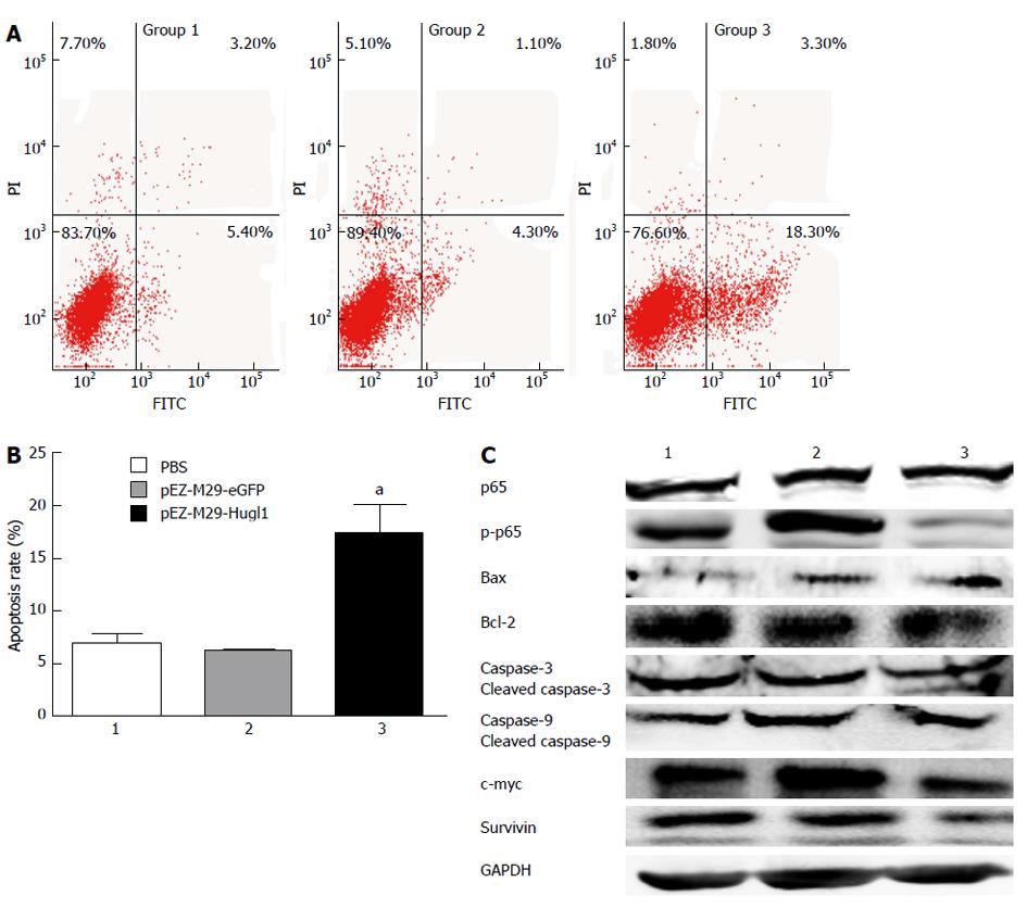
Figure 3 Effect of Hugl-1 on apoptosis of Eca109 cells in vitro.
A: Cells were treated for 48 h and were then processed for FACS by staining with Annexin V-fluorescein isothiocyanate (FITC) and propidium iodide (PI); B: After transfection with pEZ-M29-Hugl1, a significant number of cells were in an early state of apoptosis, and a population of cells had progressed to a later stage of apoptosis; C: Up-regulation of Hugl-1 led to a change of the protein levels of p65, p-p65, Bax, Bcl-2, caspase-3 and -9, survivin and c-myc among the three cell lines. All experiments were performed three times independently. aP < 0.05 vs the pEZ-M29-eGFP-treated and phosphate buffered solution (PBS)-treated groups. GAPDH: Glyceraldehyde 3-phosphate dehydrogenase.
-
Citation: Song J, Peng XL, Ji MY, Ai MH, Zhang JX, Dong WG. Hugl-1 induces apoptosis in esophageal carcinoma cells both
in vitro andin vivo . World J Gastroenterol 2013; 19(26): 4127-4136 - URL: https://www.wjgnet.com/1007-9327/full/v19/i26/4127.htm
- DOI: https://dx.doi.org/10.3748/wjg.v19.i26.4127


