Copyright
©2013 Baishideng Publishing Group Co.
World J Gastroenterol. Jul 7, 2013; 19(25): 4031-4038
Published online Jul 7, 2013. doi: 10.3748/wjg.v19.i25.4031
Published online Jul 7, 2013. doi: 10.3748/wjg.v19.i25.4031
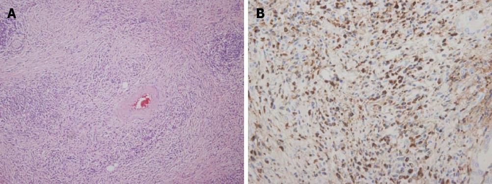
Figure 4 Histology and immunoglobulin G4 immunohistochemical staining.
A: Hematoxylin and eosin staining shows typical finding of lymphoplasmacytic sclerosing pancreatitis (× 200); B: Immunoglobulin G4 (IgG4) staining shows dense infiltration of IgG4 positive cells (× 400).
- Citation: Paik WH, Ryu JK, Park JM, Song BJ, Park JK, Kim YT, Lee K. Clinical and pathological differences between serum immunoglobulin G4-positive and -negative type 1 autoimmune pancreatitis. World J Gastroenterol 2013; 19(25): 4031-4038
- URL: https://www.wjgnet.com/1007-9327/full/v19/i25/4031.htm
- DOI: https://dx.doi.org/10.3748/wjg.v19.i25.4031


