Copyright
©2013 Baishideng Publishing Group Co.
World J Gastroenterol. Jun 28, 2013; 19(24): 3761-3769
Published online Jun 28, 2013. doi: 10.3748/wjg.v19.i24.3761
Published online Jun 28, 2013. doi: 10.3748/wjg.v19.i24.3761
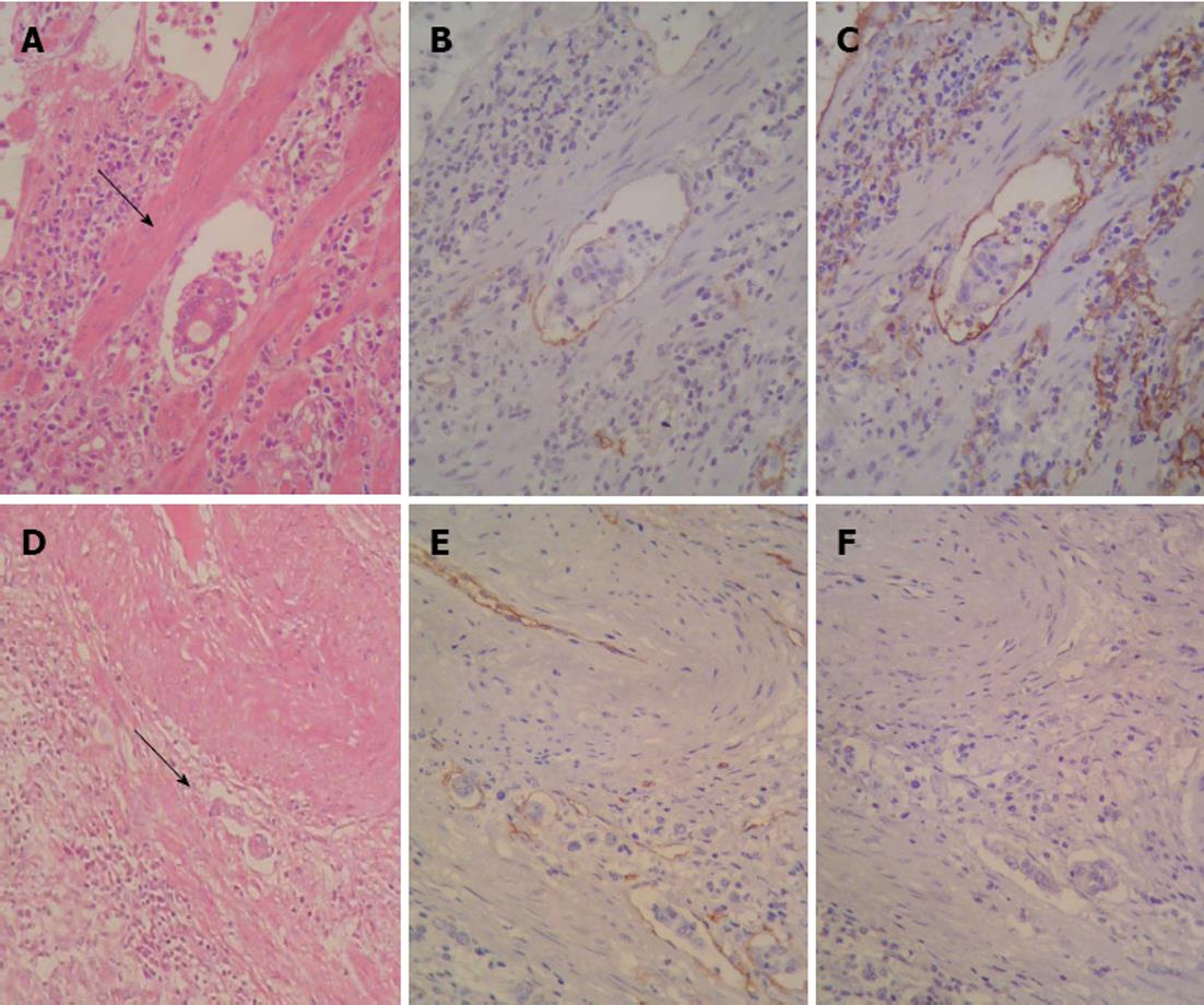
Figure 1 Sequential sections stained with hematoxylin and eosin and Immunohistochemistry showing neoplastic cell emboli within a space surrounded by the endothelial lining (arrows).
A: Lymphatic vessel invasion (LVI)-hematoxylin and eosin (HE, × 400); B: LVI CD34 (× 400); C: LVI D2-40 (× 400); D: Blood vessel invasion (BVI) (HE, × 100); E: BVI CD34 (× 200); F: BVI D2-40 (× 200).
- Citation: Gresta LT, Rodrigues-Júnior IA, Castro LPF, Cassali GD, Cabral MMD&. Assessment of vascular invasion in gastric cancer: A comparative study. World J Gastroenterol 2013; 19(24): 3761-3769
- URL: https://www.wjgnet.com/1007-9327/full/v19/i24/3761.htm
- DOI: https://dx.doi.org/10.3748/wjg.v19.i24.3761


