Copyright
©2013 Baishideng Publishing Group Co.
World J Gastroenterol. Jun 14, 2013; 19(22): 3524-3527
Published online Jun 14, 2013. doi: 10.3748/wjg.v19.i22.3524
Published online Jun 14, 2013. doi: 10.3748/wjg.v19.i22.3524
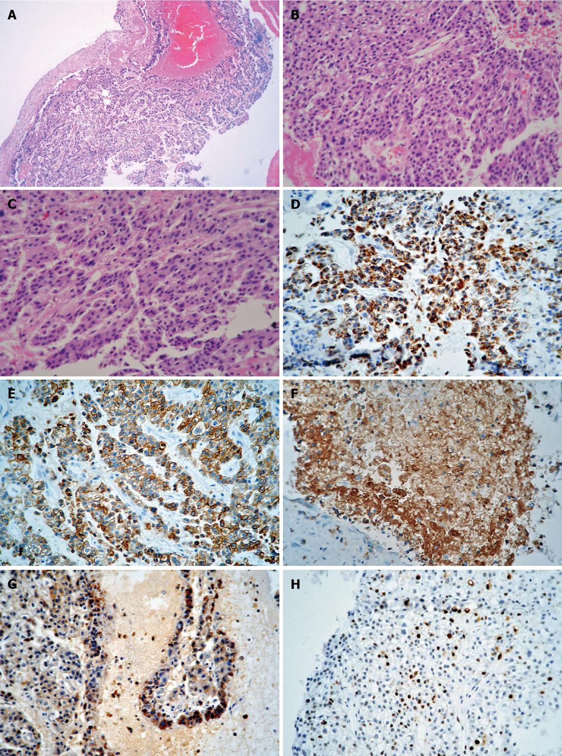
Figure 2 Histological and immunohistochemical features.
A: Low-power microphotograph showing hepatoid areas (hematoxylin and eosin; original magnification, × 100); B, C: Hepatoid adenocarcinoma composed of cells with a clear cytoplasm arranged in a nested or trabecular pattern (hematoxylin and eosin; original magnification, × 400); D-H: Intracytoplasmic expression of hepatocyte paraffin 1, cytokeratin 8/18, polyclonal carcinoembryonic antigen and S-100 as well as Ki-67-positive nuclei (original magnification, × 400).
- Citation: Wang Y, Liu YY, Han GP. Hepatoid adenocarcinoma of the extrahepatic duct. World J Gastroenterol 2013; 19(22): 3524-3527
- URL: https://www.wjgnet.com/1007-9327/full/v19/i22/3524.htm
- DOI: https://dx.doi.org/10.3748/wjg.v19.i22.3524


