Copyright
©2013 Baishideng Publishing Group Co.
World J Gastroenterol. Jun 7, 2013; 19(21): 3281-3290
Published online Jun 7, 2013. doi: 10.3748/wjg.v19.i21.3281
Published online Jun 7, 2013. doi: 10.3748/wjg.v19.i21.3281
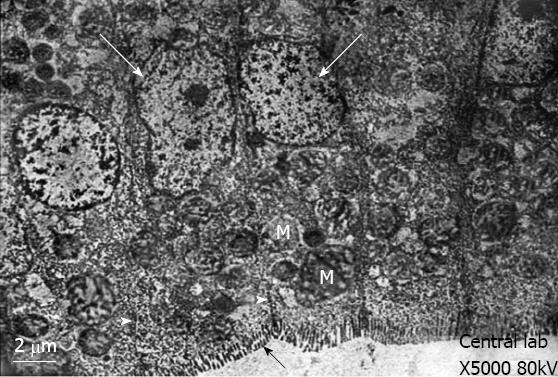
Figure 2 Transmission electron-micrograph of duodenum of rat of control group showing regularly arranged enterocytes with vesicular nucleus (white arrows) and continuous microvillus border (black arrow).
Notice the cell junction (white arrowhead) between the enterocytes and abundant mitochondria (M).
- Citation: Selim ME, Al-Ayadhi LY. Possible ameliorative effect of breastfeeding and the uptake of human colostrum against coeliac disease in autistic rats. World J Gastroenterol 2013; 19(21): 3281-3290
- URL: https://www.wjgnet.com/1007-9327/full/v19/i21/3281.htm
- DOI: https://dx.doi.org/10.3748/wjg.v19.i21.3281


