Copyright
©2013 Baishideng Publishing Group Co.
World J Gastroenterol. Jun 7, 2013; 19(21): 3263-3271
Published online Jun 7, 2013. doi: 10.3748/wjg.v19.i21.3263
Published online Jun 7, 2013. doi: 10.3748/wjg.v19.i21.3263
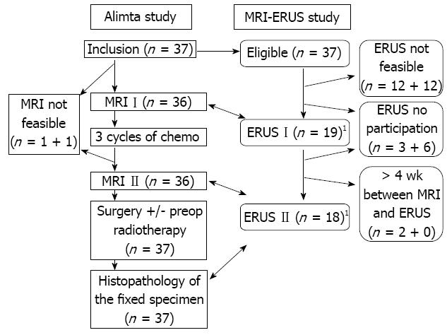
Figure 1 Study algorithm of the treatment and the examinations.
The stages and sizes using magnetic resonance imaging (MRI) were compared with the corresponding stages and sizes using endosonography (ERUS) before and after chemotherapy and with postoperative histopathology. The figure shows the assessments for size. 1Another 10 patients before chemo and 5 after chemo had complete pairs of MRI and ERUS assessments for stage.
- Citation: Swartling T, Kälebo P, Derwinger K, Gustavsson B, Kurlberg G. Stage and size using magnetic resonance imaging and endosonography in neoadjuvantly-treated rectal cancer. World J Gastroenterol 2013; 19(21): 3263-3271
- URL: https://www.wjgnet.com/1007-9327/full/v19/i21/3263.htm
- DOI: https://dx.doi.org/10.3748/wjg.v19.i21.3263


