Copyright
©2013 Baishideng Publishing Group Co.
World J Gastroenterol. Jun 7, 2013; 19(21): 3217-3225
Published online Jun 7, 2013. doi: 10.3748/wjg.v19.i21.3217
Published online Jun 7, 2013. doi: 10.3748/wjg.v19.i21.3217
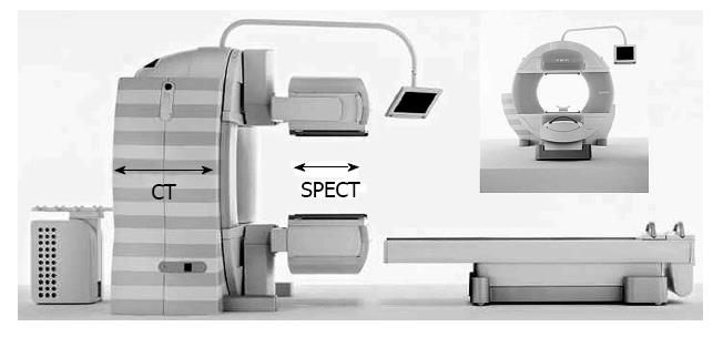
Figure 3 Integrated computed tomography and single-photon emission computed tomography system, consisting of variable angle dual-detector single-photon emission computed tomography and 6-slice computed tomography.
The system allows the seamless transition from a single-photon emission computed tomography (SPECT) examination to a computed tomography (CT) examination.
- Citation: Sumiyoshi T, Shima Y, Tokorodani R, Okabayashi T, Kozuki A, Hata Y, Noda Y, Murata Y, Nakamura T, Uka K. CT/99mTc-GSA SPECT fusion images demonstrate functional differences between the liver lobes. World J Gastroenterol 2013; 19(21): 3217-3225
- URL: https://www.wjgnet.com/1007-9327/full/v19/i21/3217.htm
- DOI: https://dx.doi.org/10.3748/wjg.v19.i21.3217


