Copyright
©2013 Baishideng Publishing Group Co.
World J Gastroenterol. Jun 7, 2013; 19(21): 3217-3225
Published online Jun 7, 2013. doi: 10.3748/wjg.v19.i21.3217
Published online Jun 7, 2013. doi: 10.3748/wjg.v19.i21.3217
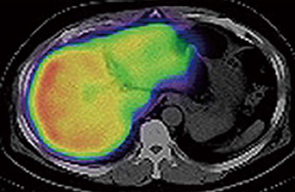
Figure 1 Computed tomography/99mTc-galactosyl human serum albumin single-photon emission computed tomography fusion image in a 58-year-old male with early gallbladder cancer.
The uptake of 99mTc-galactosyl human serum albumin was markedly decreased in the interior portion of the left lobe. This computed tomography image was at the level of the center of the left lobe, and was neither the cranial edge nor the caudal edge of that lobe.
- Citation: Sumiyoshi T, Shima Y, Tokorodani R, Okabayashi T, Kozuki A, Hata Y, Noda Y, Murata Y, Nakamura T, Uka K. CT/99mTc-GSA SPECT fusion images demonstrate functional differences between the liver lobes. World J Gastroenterol 2013; 19(21): 3217-3225
- URL: https://www.wjgnet.com/1007-9327/full/v19/i21/3217.htm
- DOI: https://dx.doi.org/10.3748/wjg.v19.i21.3217


